
Fig. S2
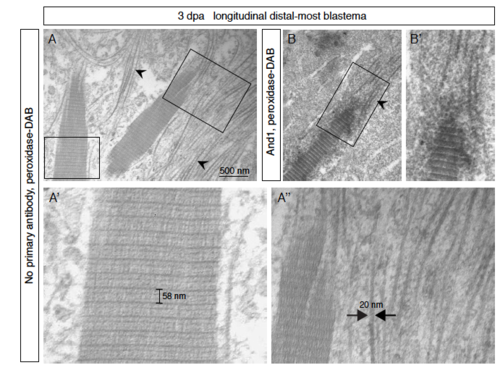
Assembly of actinotrichia detected by electron microscopy of the distal-most blastema. (A-B) Electron microscopy of the distal-most blastema at 3 dpa was performed after peroxidase- DAB immunohistochemical staining following no primary antibody (control; A) or in combination with And1 antibody (darker immunolabeled actinotrichia; B). Thin fibrils of a constant width of ≈ 20 nm (arrowheads; A’’) assemble into higher order striated fibers (A’). (A’) The main periodic striation of ≈ 60 nm is further subdivided into finer bands. (A’’) An accretion of fibrils on the extremities and surface of the actinotrichia are observed, so that the units may grow in width and in length. |
Reprinted from Developmental Biology, 433(2), König, D., Page, L., Chassot, B., Jaźwińska, A., Dynamics of actinotrichia regeneration in the adult zebrafish fin, 416-432, Copyright (2017) with permission from Elsevier. Full text @ Dev. Biol.