
Fig. 3
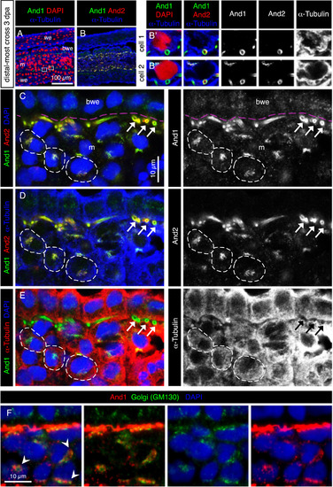
The subcellular distribution of And1 and And2 suggests their vesicular transport in mesenchymal cells. (A-F) Immunofluorescence staining of transversal sections of the distal-most outgrowth at 3 dpa (A-E) Quadruple staining for And1, And2, the cytoplasmic marker α-Tubulin and nuclear DAPI. (A, B) Overview of the tissue and magnification of two cells in the blastema. And1/2 antibodies detect ring-like structures, which are cross-sections of actinotrichia fibers. Actinotrichia are partially engulfed by mesenchymal cells, but remain extracellular. (C-E) The same specimen of the distal-most outgrowth presented in a different combination of stainings and colors. Pink dashed lines indicate the position of the basement membrane. And1 and And2 colocalize at the circumference of actinotrichia fibers (white arrows). Dotted distribution of both proteins in the α-Tubulin-labeled cytoplasm (dashed outline) indicates a subcellular localization in the vesicular system. And1 is detected in the basal wound epithelium and mesenchyme, while And2 is detected only in the mesenchyme. N = 4 fins. (F) Immunofluorescence for Golgi matrix protein GM130 (green) and And1 (red), showing colocalization of both markers in a form of perinuclear aggregates. m: mesenchyme, we: wound epidermis, bwe: basal wound epithelium. N = 4 fins. |
| Antibodies: | |
|---|---|
| Fish: | |
| Condition: | |
| Anatomical Terms: | |
| Stage: | Adult |
Reprinted from Developmental Biology, 433(2), König, D., Page, L., Chassot, B., Jaźwińska, A., Dynamics of actinotrichia regeneration in the adult zebrafish fin, 416-432, Copyright (2017) with permission from Elsevier. Full text @ Dev. Biol.