
Fig. 2-S1
- ID
- ZDB-FIG-180508-13
- Publication
- Aguillon et al., 2018 - Cell-type heterogeneity in the early zebrafish olfactory epithelium is generated from progenitors within preplacodal ectoderm
- Other Figures
- All Figure Page
- Back to All Figure Page
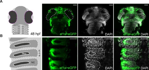
Expression of eGFP is detected throughout the head after switching of the loxP-DsRed-loxP-egfp cassette, including in known neural crest-derived lineages. (A) Low magnification images of the entire head of a double-heterozygous Tg(−28.5Sox10:Cre);Tg(ef1a:loxP-DsRed-loxP-egfp) embryo at 48 hpf labelled for eGFP (green); nuclei are labelled with DAPI (grey). The first panel shows a schematic representation of a 48 hpf embryonic head indicating the region seen in (B). eGFP is detected throughout the head. Scalebars represent 50 μm. (B) High magnification images of the embryo shown in (A). The first panel shows a schematic representation of three pharyngeal arches indicating the endodermal (End), mesodermal (Mes) and neural crest (NC) contributions. eGFP is detected specifically in the neural crest-derived contribution. Scalebars represent 20 μm. |