
Fig. 8
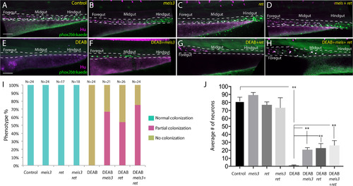
Ectopic expression of meis3 and/or ret is sufficient to partially rescue gut colonization in embryos temporally lacking RA. (A-H) Maximum intensity confocal projection images show Hu+/-8.3phox2bb:Kaede+ enteric neurons along the gut of (A) control larvae, (B) larvae expressing 40 pg of meis3, (C) larvae expressing 50 pg of ret, (D) larvae expressing 40 pg meis3 and 50 pg ret; (E) DEAB treated larvae, (F) DEAB treated larvae expressing 40 pg meis3, (G) DEAB treated larvae expressing 50 pg ret, (H) DEAB treated larvae expressing 40 pg meis3 and 50 pg ret. (E) Bar graphs depicting the percentage of larvae exhibiting normal colonization (neurons along whole length of gut), partial colonization (neurons present to the midgut) and no colonization (no neurons along the gut). (J) Bar graphs showing the average number of neurons for the rescue conditions shown in A-H. Error bars indicate S.E.M. **, p<.01 with Student's t-test. Scale bar in A-D: 60 μM. |
Reprinted from Developmental Biology, 433(1), Uribe, R.A., Hong, S.S., Bronner, M.E., Retinoic acid temporally orchestrates colonization of the gut by vagal neural crest cells, 17-32, Copyright (2017) with permission from Elsevier. Full text @ Dev. Biol.