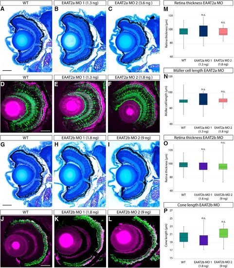
Retinal histology of EAAT2 morphant zebrafish larvae. Histologic analysis of retinal sections of WT, EAAT2a, and EAAT2b morphant zebrafish larvae (5 dpf) stained with Richardson–Romeis (A–C and G–I). Immunostaining of glutamine synthetase (green) labeling Müller glia cells counterstained with Bodipy (magenta; D–F) of WT and EAAT2a morphant (E, EAAT2a MO 1; F, EAAT2a MO 2) 5-dpf retinal sections. Anti–Zpr-1 immunostaining (labeling red and green cones, shown in green) on WT (J) and EAAT2b morphant (K, EAAT2b MO 1; L, EAAT2b MO 2) retinal sections counterstained with Bodipy (magenta). Knockdown of neither EAAT2a (B, EAAT2a MO 1; C, EAAT2a MO 2) nor EAAT2b (H, EAAT2b MO 1; I, EAAT2b MO 2) causes any defect in retinal lamination. Thickness of the retina was assessed on WT and morphant larvae and did not reveal any significant difference in the thickness of the retina in either EAAT2a or EAAT2b morphants (M, O), yielding p values of 0.997 and 0.935 for EAAT2a MO 1 and EAAT2a MO 2, respectively, and 0.658 and 0.922 for EAAT2b MO 1 and EAAT2b MO 2 (all in comparison to WT). Moreover, knockdown of EAAT2a does not significantly influence Müller glia cell length (N), nor does the loss of EAAT2b result in cone length alteration (P). Statistical analysis of the cell length yielded p values of 0.969 and 0.989 for EAAT2a MO 1 and EAAT2a MO 2, respectively, and 0.911 and 0.631 for EAAT2b MO 1 and EAAT2b MO 2 (in comparison to WT). All scale bars are 50 µm. Scale bar in A also applies to B and C; scale bar in D also applies to E and F; scale bar in G also applies to H and I; and scale bar in J also applies to K and L.
|
