
Fig. S5
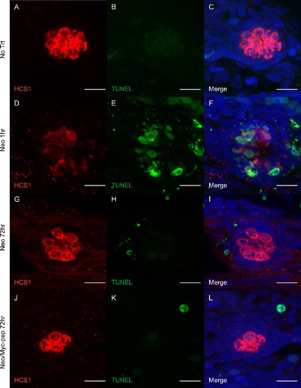
The Myc inhibitor does not induce apoptosis. 5-dpf zebrafish larvae with neomycin treatment were then treated with or without 100 nM c-MYC inhibitor Int-H1-S6A, F8A for 72 hrs (G-I, J-L). Larvae without neomycin and inhibitor treatment (No Trt) and larvae collected 1 hr after neomycin treatment were used as controls. The fish were stained with HCS1 antibody (A,D,G,J) to label HCs and TUNEL assay (B,E,H,K) to measure apoptosis. No significant difference in apoptosis signal was observed between inhibitor-treated and non-treated fish (TUNEL+ cells per neuromast: 0.4 ± 0.2 for No Trt, n = 14; 0.4 ± 0.1 for Neo 72hr, n = 15; 0.6 ± 0.2 for Neo/Myc-pep 72hr, n = 15). All TUNEL signals were from outside of the neuromast (I,L). In the positive control (D-F), a significant increase in the TUNEL+ cells were seen inside the neuromast. Scale bars: 10 μm |