
Fig. 5
- ID
- ZDB-FIG-171101-33
- Publication
- Astone et al., 2015 - A GFP-Tagged Gross Deletion on Chromosome 1 Causes Malignant Peripheral Nerve Sheath Tumors and Carcinomas in Zebrafish
- Other Figures
- All Figure Page
- Back to All Figure Page
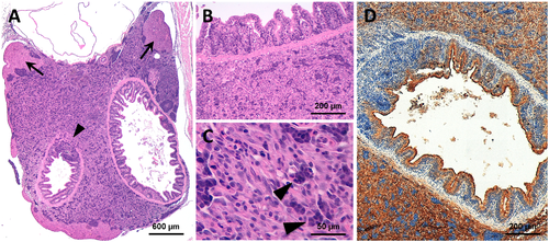
Representative histological features of the ia2 abdominal carcinomas. (A) Abdominal tumors arise beneath the swim bladder, in close proximity with the gut, pancreas and liver. The neoplastic cells occasionally infiltrate the hepatic parenchyma (arrows) and the intestinal wall (arrowhead). (B, C) Cytologically, the tumor is composed of polygonal cells with abundant eosinophilic cytoplasm. In most cases, a sub-population of epithelioid cells with more basophil cytoplasm is also faced (arrow). (D) The abdominal carcinomas consistently express the pan-cytokeratin AE1/AE3 (brown staining). See Fig 4A for the normal abdominal region. |
| Fish: | |
|---|---|
| Observed In: | |
| Stage: | Adult |