
Fig. S3
- ID
- ZDB-FIG-170921-43
- Publication
- Kara et al., 2017 - miR-27 regulates chondrogenesis by suppressing Focal Adhesion Kinase during pharyngeal arch development
- Other Figures
- All Figure Page
- Back to All Figure Page
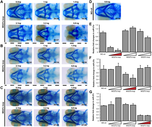
Dose-dependent changes in cartilage defects by mature miR-27 a, b and cknock-down. Head cartilage staining of 4 dpf old embryos injected with different doses of the MO-27a loop (A) MO-27b loop (B) MO-27c loop (C) morpholinos complementary to the loop and Dicer cleavage site of the corresponding precursor miR-27. All are ventral views and the ratio of the observed phenotype to the total number of embryos is shown for each image. (D) Alcian blue staining of the 4dpf embryos injected with 4.8 ng MO-ctl. qRT-PCR of miR-27a (E), miR-27b (F), and miR-27c (G) in 30hpf old embryos injected with 4.8 ng MO-ctl, MO27a loop, MO27b loop, and MO27c loop at either 2.4 ng or 4.8 ng. |
Reprinted from Developmental Biology, 429(1), Kara, N., Wei, C., Commanday, A.C., Patton, J.G., miR-27 regulates chondrogenesis by suppressing Focal Adhesion Kinase during pharyngeal arch development, 321-334, Copyright (2017) with permission from Elsevier. Full text @ Dev. Biol.