
Fig. 1
- ID
- ZDB-FIG-161107-4
- Publication
- Marín-Juez et al., 2016 - Fast revascularization of the injured area is essential to support zebrafish heart regeneration
- Other Figures
- All Figure Page
- Back to All Figure Page
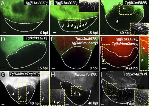
The zebrafish heart displays fast revascularization of the damaged area. (A-C) Tg(fli1a:EGFP) ventricles at 0, 15, and 40 hpi. White arrowheads point to coronary vessel sprouts in the injured area; asterisk marks an area of coronary plexus (n = 4). Insets show high-magnification images of early coronary sprouts (B) and the coronary plexus (C). (D) Tg(kdrl:EGFP) heart at 15 hpi (n = 3). (E and F) Tg(fli1a:EGFP);Tg(kdrl:mCherry) ventricles extracted at 0 hpi and cultured in explant medium for 24 h (0+24 hpi) (n = 3). White arrowhead points to kdrl:EGFP+ vessels sprouting into the injured area. Inset shows overlay image of a sprouting coronary vessel. White arrows point to fli1a:EGFP+/kdrl:mCherry+ coronary vessel sprouting into the injured area. (G) Tg(Dll4in3:TagRFP) ventricle at 40 hpi (n = 5). Inset shows a Dll4in3:TagRFP+ sprouting coronary artery. (H and I) Tg(cxcr4a:YFP) ventricles at 40 hpi (n = 3) and 7 dpi (n = 4). Insets show cxcr4a:YFP+ sprouting coronaries; white arrowheads point to coronary arteries sprouting into the injured area. Dotted lines delineate the injured area. (Scale bars: 50 µm.) |
| Genes: | |
|---|---|
| Fish: | |
| Condition: | |
| Anatomical Terms: | |
| Stage: | Adult |