
FIGURE
Fig. S2
- ID
- ZDB-FIG-161107-6
- Publication
- Marín-Juez et al., 2016 - Fast revascularization of the injured area is essential to support zebrafish heart regeneration
- Other Figures
- All Figure Page
- Back to All Figure Page
Fig. S2
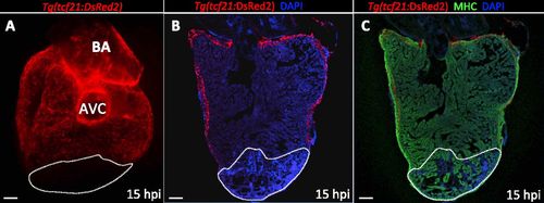
No epicardial cells are found in the damaged area at 15 hpi. (A) Dorsal view of a 15 hpi Tg(tcf21:DsRed2) ventricle (n = 3). (B and C) Sections of a Tg(tcf21:DsRed2) ventricle at 15 hpi (n = 3). Cardiomyocytes are immunostained with anti-MHC antibody (red), and nuclei are counterstained with DAPI (blue). White dotted lines delineate the injured area. AVC, atrioventricular canal; BA, bulbus arteriosus. (Scale bars: 100 µm.) |
Expression Data
Expression Detail
Antibody Labeling
Phenotype Data
Phenotype Detail
Acknowledgments
This image is the copyrighted work of the attributed author or publisher, and
ZFIN has permission only to display this image to its users.
Additional permissions should be obtained from the applicable author or publisher of the image.
Full text @ Proc. Natl. Acad. Sci. USA