
FIGURE
Fig. 4
- ID
- ZDB-FIG-160909-10
- Publication
- Anthony et al., 2016 - An Optimized Small Tissue Handling System for Immunohistochemistry and In Situ Hybridization
- Other Figures
- All Figure Page
- Back to All Figure Page
Fig. 4
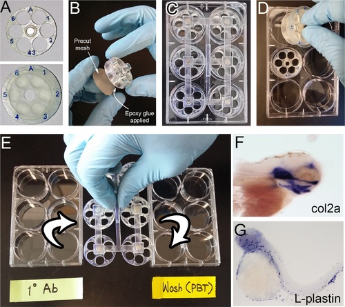
Actual photos of the system and its use for in situ hybridization. (A) Enhanced labeling with artist’s modeling clay and coating. (B) Mesh attachment with epoxy glue. (C) A fully loaded plate with a magnetic lifter attached. (D) With only a few samples, individual chambers can be conveniently transferred manually. Chamber A, with its lid closed, was picked up by hand, whereas the chamber B is shown without its lid in pace. (E) 6 chambers (36 samples) are handled simultaneously with a magnetic lifter. (F, G) Examples of whole mount in situ hybridization results. |
Expression Data
| Genes: | |
|---|---|
| Fish: | |
| Anatomical Terms: | |
| Stage Range: | 20-25 somites to Day 5 |
Expression Detail
Antibody Labeling
Phenotype Data
Phenotype Detail
Acknowledgments
This image is the copyrighted work of the attributed author or publisher, and
ZFIN has permission only to display this image to its users.
Additional permissions should be obtained from the applicable author or publisher of the image.
Full text @ PLoS One