
Fig. 4
- ID
- ZDB-FIG-160817-45
- Publication
- Naylor et al., 2016 - BMP and retinoic acid regulate anterior-posterior patterning of the non-axial mesoderm across the dorsal-ventral axis
- Other Figures
- All Figure Page
- Back to All Figure Page
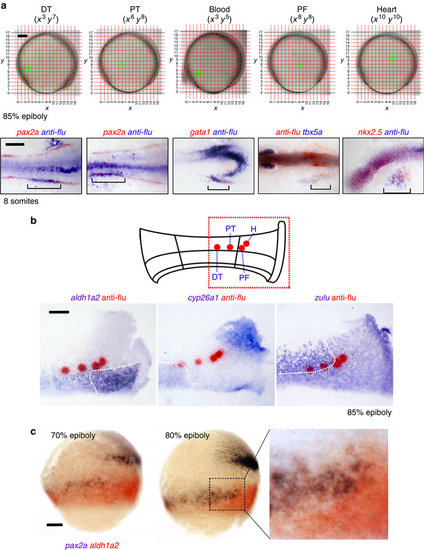
Fate-mapping analysis of late gastrula embryos show proximal tubule and pectoral fin progenitors are juxtaposed to the aldh1a2high domain. (a) Cells on the left-hand side of embryos at the 85% epiboly stage were lineage-labelled at various co-ordinates on the Cartesian grid shown (embryos are orientated with animal pole towards the top and dorsal side to the right). At the 10-somites stage, co-localization of uncaged cells and kidney (pax2a+), blood (gata1+), pectoral fin (tbx5a+) and heart (nkx2.5+) tissues was determined by whole-mount double in situ hybridization. Labelled cells on the right-hand side of the embryo (white asterisk) are the result of the laser passing through the embryo and uncaging cells on the contralateral side. Embryos are flat-mounted with anterior to the left (note: tbx5a is stained purple due to being a weaker probe). (b) Top schematic shows a flat-mounted embryo with the highlighted region corresponding to the panels below and the positions that label progenitors of the proximal (PT) and distal (DT) tubules, pectoral fin (PF) and heart (H). Bottom panels show whole-mount double in situ hybridization of these uncaged populations (red) relative to the expression domains of aldh1a2, cyp26a1 and zulu (purple) at 85% epiboly. (c) Lateral views of 70% (left) and 80% (centre) epiboly-stage embryos double stained for aldh1a2 (purple/black) and pax2a (red) transcripts. Higher magnified view of the indicated region is shown in the right-hand panel. A, animal pole; V, vegetal pole. Schematic at the bottom represents an outline of PLM. IM, intermediate mesoderm; LPM, lateral plate mesoderm. Scale bar in a, 100 µm in top panels and 200 µm in lower panels. Scale bars in b and c, 100 µm. |