
Fig. 3
- ID
- ZDB-FIG-160816-4
- Publication
- Venero Galanternik et al., 2015 - Heparan Sulfate Proteoglycans Regulate Fgf Signaling and Cell Polarity during Collective Cell Migration
- Other Figures
- All Figure Page
- Back to All Figure Page
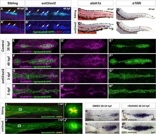
Fgf Signal Transduction Is Progressively Lost in extl3/ext2 Mutant Prim (A-B′) ZO-1 staining reveals apical constrictions (white arrows) in 48 hpf extl3/ext2 neuromasts and prim rosettes, but not in interneuromast clumps (yellow arrow). (C-D′) LL hair cells precursors are present in 48 hpf extl3/ext2 mutants, as evidenced by atoh1a and s100t expression. Black arrows in (C), (D), and (D′) indicate the position of the prim. (E-H′′) Dp-ERK staining reveals remaining low levels of Fgf signaling in the 48 hpf prim. Dp-ERK is progressively downregulated as rosettes collapse (E′′-H′′). By 5 dpf, no protein remains inside the prim (H′). (I-J′) By 3 dpf, most of the formed hair cells die inside the collapsing neuromast. (I′ and J′) Neuromasts outlined in the white boxes in (I) and (J). (K-L′) Inhibition of Fgf signaling accelerates the onset of the extl3/ext2 phenotype. extl3/ext2 mutants and their siblings treated with SU5402 from 28 to 34 hpf show complete upregulation of the Wnt target lef1. See also Figures S2, S3, and S5 and Movie S4. |
| Genes: | |
|---|---|
| Antibody: | |
| Fish: | |
| Condition: | |
| Anatomical Terms: | |
| Stage Range: | Prim-15 to Long-pec |
| Fish: | |
|---|---|
| Condition: | |
| Observed In: | |
| Stage Range: | Prim-15 to Protruding-mouth |