
FIGURE
Fig. S3
- ID
- ZDB-FIG-160714-24
- Publication
- Richardson et al., 2016 - Re-epithelialization of cutaneous wounds in adult zebrafish uses a combination of mechanisms at play during wound closure in embryonic and adult mammals
- Other Figures
- All Figure Page
- Back to All Figure Page
Fig. S3
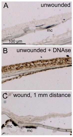
The epidermis around closing full-thickness wounds lacks apoptotic keratinocytes. (A-C) TUNEL staining on sections through unwounded (A.B) and wounded (C) trunk region, 4 hpw, with (B) and without (A,C) DNase treatment to induce DNA fragmentation, as formerly described (Fischer et al., 2014). No TUNEL-positive cells are detectable in the epidermis 1 mm from the wound, indicating that the thinning of the epidermis in this region is not due to keratinocyte apoptosis. Similar results were obtained via immunofluorescent labelling of activated caspase 3 (data not shown). Abbreviation: mc, melanocyte. |
Expression Data
Expression Detail
Antibody Labeling
Phenotype Data
Phenotype Detail
Acknowledgments
This image is the copyrighted work of the attributed author or publisher, and
ZFIN has permission only to display this image to its users.
Additional permissions should be obtained from the applicable author or publisher of the image.
Full text @ Development