
Fig. 4
- ID
- ZDB-FIG-160714-19
- Publication
- Richardson et al., 2016 - Re-epithelialization of cutaneous wounds in adult zebrafish uses a combination of mechanisms at play during wound closure in embryonic and adult mammals
- Other Figures
- All Figure Page
- Back to All Figure Page
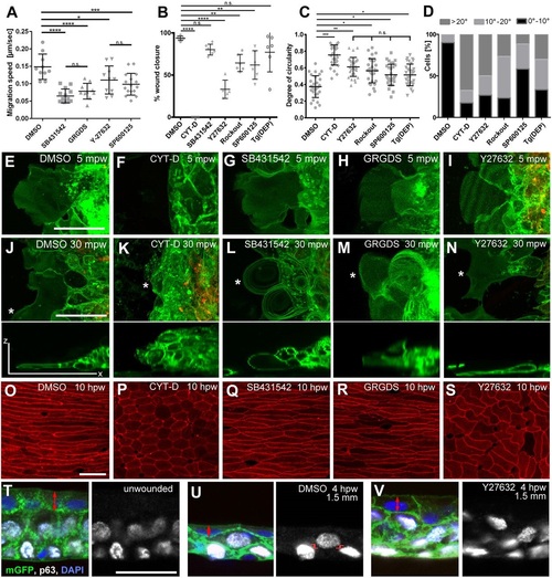
Re-epithelialization requires TGF-β/GRGDS-dependent keratinocyte crawling at the LE and Rock/JNK-dependent epithelial morphogenesis in the following epidermis. (A-D) Quantification of effects caused by the indicated inhibitors on LE migration speeds in partial-thickness wounds (A), and on the extent of wound closure (B), the degree of circularity (C) and the orientation of the long axis (D) of adjacent superficial cells of full-thickness wounds at 10hpw. Tg(DEP), transgenic inhibition of PCP. In A-C, mean values and s.d. are indicated. Values in A were determined from time-lapse movies (e.g. Movies 2-5; n≥5 per condition), values in B from Methylene Blue penetration assays as shown in Fig. S4F,G, and values in C,D from images as shown in O-S. *P<0.05; **P<0.01; ***P<0.005; ****P<0.001; n.s., not significant; one-way ANOVA with a Dunnett′s post-hoc test. (E-N) Live confocal images of the LE of Tg(actb2:hras-egfp), Tg(krt4:mCherry) double transgenic fish at 5min (E-I) and 30min (J-N) after partial-thickness wounding (mpw), treated with indicated inhibitors. Lower panels of J-N show z-projections of the LE cell marked by the asterisk in the panels above. At 5mpw, all cases except the CYT-D treatment display normal protrusive activity at the LE, whereas at 30mpw, SB431542- and GRGDS-treated LE keratinocytes display roundish, shorter and thicker lamellipodia, indicative of lamellipodial retraction. (O-S) Phalloidin staining of the adjacent epidermis of full-thickness wounds at 10hpw demonstrating compromised and uncoordinated cell elongation upon treatment with CYT-D (P) and Y27632 (S), but not SB431542 (Q) or GRGDS (S). (T-V) p63, GFP double immunofluorescence and DAPI labelling of sections through Tg(actb2:hras-egfp) fish at indicated conditions and distances from full-thickness wounds. Left panels, merge; right panels, p63 channels. Y27632-treated wound (V) displays reduced flattening of superficial cells (double-headed red arrows) and reduced frequency of radial intercalations, characterized by a partial overlap of the positions along the epidermal apical-basal axis occupied by adjacent basal and suprabasal p63+ nuclei (marked in U by red lines). Quantifications from three individual fish per condition (1-1.5mm wound distance for U,V) yielded the following frequencies (no. of partially overlapping p63+ nuclei/total no. of p63+ nuclei). Unwounded in T, 6.1±5.3%; DMSO-treated wound in U, 51.3±3.7%; Y27632-treated wound in V, 17.2±4.9%. P=0.00027557 (T versus U); P=0.056567873 (T versus V); P=0.000651499 (U versus V); Student′s t-test. Scale bars: 20µm. |
| Gene: | |
|---|---|
| Fish: | |
| Conditions: | |
| Anatomical Term: | |
| Stage: | Adult |