
Fig. 5
- ID
- ZDB-FIG-160316-25
- Publication
- Xu et al., 2016 - Four and a Half LIM Domains 1b (Fhl1b) Is Essential for Regulating the Liver versus Pancreas Fate Decision and for β-Cell Regeneration
- Other Figures
- All Figure Page
- Back to All Figure Page
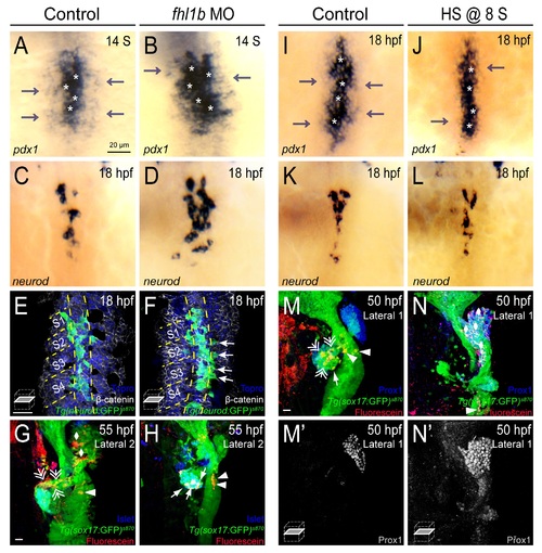
Fhl1b regulates the patterning and subsequent fate of the medial and lateral endodermal progenitors. (A-D) Whole-mount in situ hybridization showing the expression of pdx1 (A and B) and neurod (C and D), comparing that of control embryos (A and C) and fhl1b morphants (B and D) at the 14-somite stage (A and B) and 18 hpf (C and D). pdx1 is expressed at high levels in the most medial cells (white asterisks) and at low levels in the lateral cells (gray arrows). neurod is expressed in the high-level pdx1-expressing cells. In fhl1b morphants, high levels of pdx1 (white asterisks) and neurod expression were expanded laterally (B and D). (E-F) Ventral confocal images showing TgBAC(neurod:EGFP)nl1, β-catenin (white), and Topro (blue) at 18 hpf (the notochord is outlined by yellow dashed lines). Somites are numbered from anterior to posterior (S1-S4). (E) In control embryos, TgBAC(neurod:EGFP)nl1-expressing cells are located close to the notochord. (F) Ectopic TgBAC(neurod:EGFP)nl1-expressing cells were found in lateral endodermal regions in fhl1b morphants (white arrows). (G and H) Confocal images of Tg(sox17:GFP)s870 embryos at 55 hpf, stained for uncaged-Fluorescein (red) and Islet (blue). In control embryos (G), lateral 2 (L2) cells gave rise to the liver (white rhombi), intestine (white arrowhead), and exocrine pancreas (white double arrows), but rarely gave rise to the endocrine pancreas. In fhl1b morphants (H), L2 cells contributed to the Islet-positive pancreatic endocrine cells (white arrows), but not to the liver or exocrine pancreas. (I-L) Whole-mount in situ hybridization showing the expression of pdx1 (I and J) and neurod (K and L) at 18 hpf, comparing control embryos (I and K) and fhl1b-overexpressing embryos (J and L, heat shock applied at the 8-somite stage). In embryos induced to overexpress fhl1b at the 8-somite stage (J and L), neurod and high levels of pdx1 expression (white asterisks in J) were maintained, while low levels of pdx1 expression (gray arrows) were reduced. (M-N′) Confocal images of Tg(sox17:GFP)s870 embryos at 50 hpf, stained for uncaged-Fluorescein (red) and Prox1 (blue in M and N; grey in M′ and N′). In control embryos (M and M′), lateral 1 (L1) cells gave rise to the exocrine pancreas (white double arrows) and the intestine (white arrowheads), but not to the liver. In embryos induced to overexpress fhl1b at the 8-somite stage (N and N′), L1 cells mostly contributed to the Prox1-positive liver cells (white rhombi). A-D and I-L, dorsal views, anterior to the top. G-H and M-N, confocal projection images; E-F, M′, and N′, confocal single-plane images, ventral views, anterior to the top. Scale bars, 20 µm. |
| Genes: | |
|---|---|
| Antibody: | |
| Fish: | |
| Condition: | |
| Knockdown Reagents: | |
| Anatomical Terms: | |
| Stage Range: | 14-19 somites to Long-pec |
| Fish: | |
|---|---|
| Condition: | |
| Knockdown Reagents: | |
| Observed In: | |
| Stage: | 14-19 somites |