
FIGURE
Fig. 6
- ID
- ZDB-FIG-160303-20
- Publication
- Bensimon-Brito et al., 2016 - Revisiting in vivo staining with alizarin red S - a valuable approach to analyse zebrafish skeletal mineralization during development and regeneration
- Other Figures
- All Figure Page
- Back to All Figure Page
Fig. 6
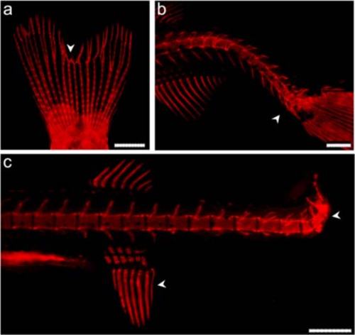
Detection of skeletal malformations in zebrafish. Deformed bony structures in a caudal fin rays and b-c different regions of the vertebral column. All regions display affected structures with different degrees of severity. White arrowheads show sites of malformation. Scale bars (a) = 2 mm; (b-c) = 0.4 mm |
Expression Data
Expression Detail
Antibody Labeling
Phenotype Data
Phenotype Detail
Acknowledgments
This image is the copyrighted work of the attributed author or publisher, and
ZFIN has permission only to display this image to its users.
Additional permissions should be obtained from the applicable author or publisher of the image.
Full text @ BMC Dev. Biol.