
FIGURE
Fig. 4
- ID
- ZDB-FIG-160303-18
- Publication
- Bensimon-Brito et al., 2016 - Revisiting in vivo staining with alizarin red S - a valuable approach to analyse zebrafish skeletal mineralization during development and regeneration
- Other Figures
- All Figure Page
- Back to All Figure Page
Fig. 4
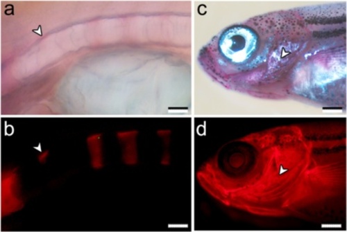
ARS staining of fixed zebrafish samples. Panels a-b show a vertebral column of a 10 dpf larva stained with 0.01 % ARS in 70 % ethanol. a Bright field observation provides less detail of the early mineralization deposits than b fluorescence observation (arrowheads). Panels c-d show cranial structures of a juvenile (30 dpf, 8 mm TL) stained with 0.01 % ARS and observed under c bright field and d fluorescent light, evidencing the higher power of detection of, e.g., the operculum (arrowheads) under fluorescent conditions. Scale bars (a, b) = 0.04 mm; (c, d) = 0.2 mm |
Expression Data
Expression Detail
Antibody Labeling
Phenotype Data
Phenotype Detail
Acknowledgments
This image is the copyrighted work of the attributed author or publisher, and
ZFIN has permission only to display this image to its users.
Additional permissions should be obtained from the applicable author or publisher of the image.
Full text @ BMC Dev. Biol.