
Fig. 7
- ID
- ZDB-FIG-150929-42
- Publication
- Rajaram et al., 2014 - Technical brief: Constant intense light exposure to lesion and initiate regeneration in normally pigmented zebrafish
- Other Figures
- All Figure Page
- Back to All Figure Page
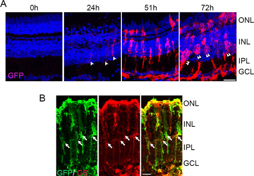
Light damage triggers tuba1a:GFP transgene expression in Müller glia (MG). A: tg:1016tuba1a:GFP zebrafish were exposed to the constant intense light paradigm, and retinas were collected at the indicated time points and assessed for GFP expression (red) by immunohistochemistry. GFP+ cells began to appear in the INL by 24 h (arrowheads). By 72 h, clusters of GFP+ cells were detected in the INL and ONL (double arrowheads). B: Immunohistochemistry for GFP and GS in 51-h light-exposed 1016tuba1a:GFP retinas. Arrows indicate GS+ and GFP+ MG. ONL, outer nuclear layer; INL, inner nuclear layer; GCL, ganglion cell layer; IPL, inner plexiform layer. Scale bars are 50 µm. |
| Gene: | |
|---|---|
| Fish: | |
| Condition: | |
| Anatomical Term: | |
| Stage: | Adult |