
Fig. 6
- ID
- ZDB-FIG-150929-41
- Publication
- Rajaram et al., 2014 - Technical brief: Constant intense light exposure to lesion and initiate regeneration in normally pigmented zebrafish
- Other Figures
- All Figure Page
- Back to All Figure Page
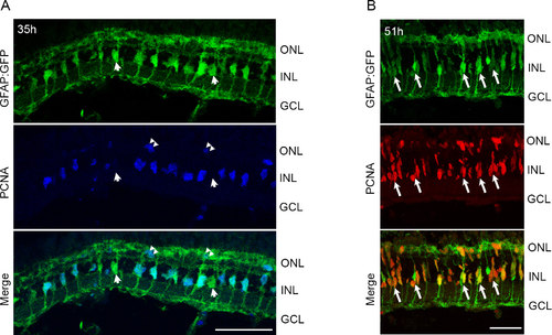
Light treatment of pigmented retinas stimulates proliferation of MG and progenitor cells. tg:GFAP:GFP fish were exposed to intense light for 35 h (A) or 51 h (B), and retinas were processed for immunohistochemistry. A: Proliferating cell nuclear antigen (PCNA) immunohistochemistry (blue) reveals co-staining with GFP+ MG in the central-dorsal retina. Few MG do not express PCNA (arrowheads in A). PCNA also labels rod precursors in the ONL (double arrowheads). B: Most GFP+ MG continue to express PCNA. Arrows indicate MG associated with small PCNA+ progenitor clusters. Few PCNA+ cells are migrating to the ONL. ONL, outer nuclear layer; INL, inner nuclear layer; GCL, ganglion cell layer. Scale bars are 50 µm. |
| Gene: | |
|---|---|
| Fish: | |
| Condition: | |
| Anatomical Term: | |
| Stage: | Adult |