
Fig. 6
- ID
- ZDB-FIG-150526-19
- Publication
- Burrows et al., 2015 - An In Vivo Requirement for the Mediator Subunit Med14 in the Maintenance of Stem Cell Populations
- Other Figures
- All Figure Page
- Back to All Figure Page
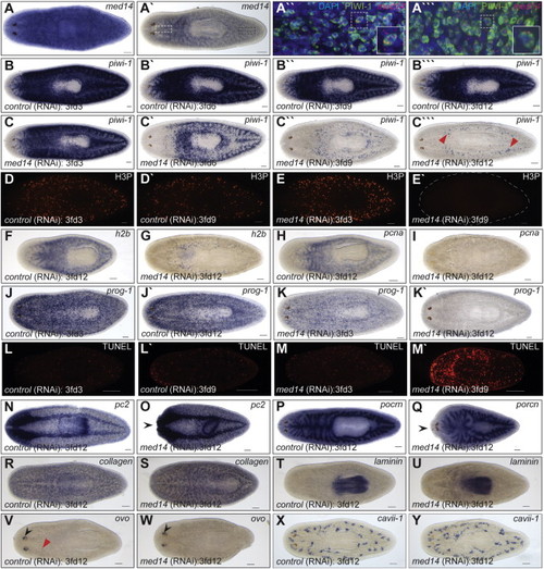
Smed-med14 Is Necessary for the Maintenance of Adult Stem Cells (A) ISH for med14 in wild-type intact animals showing ubiquitous staining. (A′) A stem cell like expression pattern is evident with reduced staining. (A′′ and A′′′) Confocal image at 25× magnification of med14 fluorescent RNA ISH (red) and PIWI antibody staining (green) in the planarian head (white dashed box in A2) and tail (black dashed box in A′) respectively. The boxed area in each is enlarged for clarification. med14 is expression in, but not limited to, the stem cell population. (B–C′′′) ISH analysis using a stem cell specific riboprobe (piwi-1) during a time course of med14(RNAi). By 3fd12, the stem cell population is largely absent in med14(RNAi) animals. The remaining piwi-1+ cells at 3fd12 (C′′′) may represent primordial germ cells (red arrowheads in M). (D–E′) Loss of proliferative phosphorylated histone H3 (H3P) +’ve cells in med14(RNAi) animals by 3fd9 (E′). (F–I) Expression of S-phase markers h2b and pcna in WT and med14 RNAi animals at 3fd12. (J–K′) By 3fd3, the progenitor cell population in med14(RNAi) animals (marked by prog-1 expression) is reduced compared with controls and completely absent by 3fd12. (L–M′) Increased cell death by 3fd9 as observed by whole-mount TUNEL analysis in med14(RNAi) animals. (N–Y) Normal expression of markers of differentiated cell types in med14(RNAi) animals as evident for the nervous system (pc2), gut (porcn), muscle (collagen), pharynx (laminin), eyes (ovo), and protonephridia (cavii-1). Head regression is evident in some treated worms (black arrow heads in O and Q). Eye progenitors (red arrow head in V) are not observed in med14(RNAi) animals despite ovo expression in the eye spots (black arrow heads in V and W). Scale bars, 100 µm. See also Figure S2. |