
Fig. S8
- ID
- ZDB-FIG-141007-86
- Publication
- Pillai-Kastoori et al., 2014 - Sox11 Is Required to Maintain Proper Levels of Hedgehog Signaling during Vertebrate Ocular Morphogenesis
- Other Figures
- All Figure Page
- Back to All Figure Page
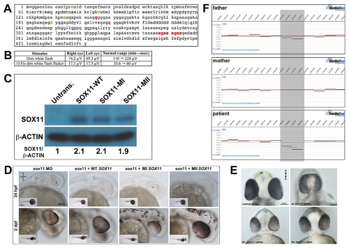
Association of SOX11 locus with ocular abnormalities. (A) Amino acid sequence of human SOX11, with previously identified non-synonomous SNPs highlighted in green. The two variants identified in the MAC patients (positions indicated in red) are novel. (B) Scotopic ERG analysis of the proband′s mother carrying the S315–354dup variant, demonstrating a reduction in the b-wave amplitude. (C) Western blot for SOX11 and β-actin in COS-7 cells transfected with SOX11 expression constructs. Densitometric analysis was performed with ImageJ software. (D) Representative brightfield images of sox11 morphants co-injected with either WT, MI (G145C), or MII (S315–354dup) SOX11 mRNA at 24 hpf and 2 dpf, taken from the set of embryos analyzed in Figure 8D. (E) Representative brightfield images of embryos overexpressing human WT, MI, or MII SOX11 mRNA, taken from the set of embryos analyzed in Figure 8F. (F) Array CGH analysis of a proband with optic nerve agenesis and microphthalmia and her parents, confirming the presence of a de novo interstitial deletion at chromosome 2p25.2 (shaded gray). D, dorsal; V, ventral; A, anterior; P, posterior; MO, morpholino; hpf, hours post fertilization; dpf, days post fertilization; L, lens. |