
Fig. S3
- ID
- ZDB-FIG-141007-82
- Publication
- Pillai-Kastoori et al., 2014 - Sox11 Is Required to Maintain Proper Levels of Hedgehog Signaling during Vertebrate Ocular Morphogenesis
- Other Figures
- All Figure Page
- Back to All Figure Page
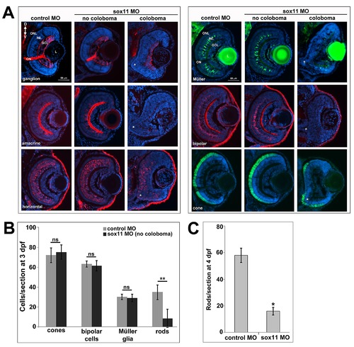
Retinal neurogenesis in sox11 morphants. (A) Retinal cell types were visualized by immunohistochemistry (ganglion, amacrine, horizontal, and bipolar cells) or with fluorescent reporter transgenic lines (Tg(gfap:GFP)mi2001 for Müller glia and Tg(3.2TαC-EGFP) for cones) in controls (left) and sox11 morphants (center, right) at 3 dpf. In sox11 morphants without coloboma (center), the retinas are well laminated and had normal numbers of ganglion, amacrine, horizontal, and bipolar cells, cone photoreceptors, and Müller glia. However, sox11 morphants with coloboma (asterisk; right) had poorly laminated retinas and reduced numbers of differentiated retinal cell types, indicating delayed retinal development. (B) Quantification of numbers of late-born retinal cell types in control and sox11 morphants without coloboma. Only rod photoreceptors displayed a significant reduction. Number of embryos analyzed: control MO, n = 19; sox11 MO without coloboma, n = 25, 3 independent repeats.**p<0.00001; ns = p>0.05, Student′s t-test.(C) At 4 dpf, sox11 morphants have more mature rod photoreceptors than at 3 dpf but the number remains significantly less than controls (*p<0.001, Student′s t-test); MO, morpholino; dpf; days post fertilization; L, lens; GCL, ganglion cell layer; INL, inner nuclear layer; ONL, outer nuclear layer; ON, optic nerve. |