
Fig. S4
- ID
- ZDB-FIG-140822-19
- Publication
- Whitesell et al., 2014 - An alpha-smooth muscle actin (acta2/alphasma) zebrafish transgenic line marking vascular mural cells and visceral smooth muscle cells
- Other Figures
- All Figure Page
- Back to All Figure Page
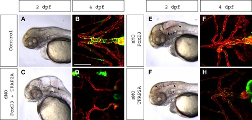
Single or double knockdown of FoxD3 or TFAP2a to block neural crest specification results in a reduction in acta2:GFP cells, but also severe ventral head and blood vessel patterning defects. Representative brightfield images of 2 dpf zebrafish embryos show that both double knockdown (dMO) of FoxD3 and TFAP2A (C) or single knockdown (sMO) of FoxD3 (E) or TFAP2A (G), results in hemorrhage which is not present in control (A). Hydrocephalus of the hindbrain ventricle is also observed in dMO and sMO FoxD3. At 4 dpf, confocal microscopy shows that the control has a well-defined heart outflow tract, with mural cell coverage (kdrl:mCherry – red vessels; acta2:EGFP – green mural cells) (B). In dMO there are severe vessel malformations and a reduction in mural cell coverage (D). In the single FoxD3 (F) and TFAP2A (H) morphants, there are also malformations and reduced mural cell coverage, although these are less severe than the double morphant. Scale bar for A, C, E, G represents 200 µm. Scale bar for B, D, F, H represents 100 μm. |