
Fig. 8
- ID
- ZDB-FIG-140822-14
- Publication
- Whitesell et al., 2014 - An alpha-smooth muscle actin (acta2/alphasma) zebrafish transgenic line marking vascular mural cells and visceral smooth muscle cells
- Other Figures
- All Figure Page
- Back to All Figure Page
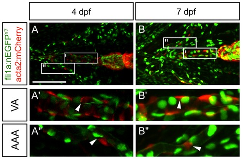
Lack of co-localization of mural cell and the ectomesenchymal neural crest marker Fli1a. Confocal images of 4 and 7(acta2:mCherry) and nuclear neural crest marker (fli1a:nEGFPy7) using ventrally staged embryos. (A) Mural cell and neural crest markers are expressed along the ventral aorta (A′) and aortic arch artery region (A′′) of the 4 dpf embryo. (B) Mural cell and neural crest markers are expressed along the ventral aorta (B′) and aortic arch region (B′′) at 7 dpf. There appears to be little to no co-localization of fluorescent markers at both 4 and 7 dpf. Scale bar in A represents 100 μm. Insets (A′, A′′, B′, B′′) are 100 μm in length. VA = Ventral Aorta, AAA = Aortic Arch Arteries. Arrowheads depict cells that no do not co-localize. |