
Fig. 5
- ID
- ZDB-FIG-140806-9
- Publication
- Teng et al., 2013 - Evaluating human cancer cell metastasis in zebrafish
- Other Figures
- All Figure Page
- Back to All Figure Page
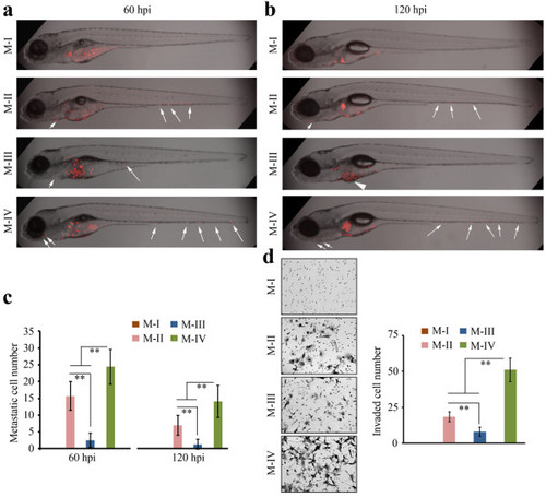
Relative metastatic potential in human cancer cells during progression from low to high grade tumors. When immortal, non-transformed MCF10A cells (M-I) were introduced into the zebrafish metastasis model (a), no spread was seen in any of the fish analyzed at 60 hpi. The cells (M-II) recovered from low grade hyperplastic lesions in mice from MCF10AT cells (MCF10A cells transformed with activated HRAS) show increased metastatic potential (arrows). M-III cells, which were derived from well differentiated carcinomas derived from M-II cells, show reduced metastatic potential, whereas M-IV cells, recovered from aggressive adenocarcinomas derived from M-II cells, show enhanced metastatic potential. The same relationship was seen at 120 hpi (b) (shown as z-stack images using 40× magnification). Quantitation of metastatic cells throughout the fish body using Fiji (c) confirms the relationship between tumor grade and metastatic potential in fish (left). At 120 hpi, although the relative proportion of metastatic cells is seen in the M-II/M-III/M-IV comparison, the overall number of cells in each tumor grade was reduced. When the same comparison was performed using the in vitro invasion assay (d), the relative proportion of invading cells in the different sub groups was again maintained. Data are presented as the mean of three independent experiments (n = 3) ± SEM, **P < 0.01. |