
Fig. S1
- ID
- ZDB-FIG-140624-31
- Publication
- Zeng et al., 2014 - Cadm4 Restricts the Production of Cardiac Outflow Tract Progenitor Cells
- Other Figures
- All Figure Page
- Back to All Figure Page
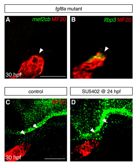
Fgf signaling influences gene expression in the vicinity of the arterial pole. (A,B) Fluorescent in situ hybridization examines expression of mef2cb (green, A) and ltbp3 (green, B) in the area adjacent to the arterial pole of the differentiated heart tube (MF20, red) at 30 hpf; images are partial confocal reconstructions. Embryos homozygous for a loss-of-function mutation in fgf8a (Reifers et al., 2000) display striking reductions in the expression of mef2cb (arrowhead, A) and ltbp3 (arrowhead, B), in comparison to the expression levels seen in wild-type embryos (compare with Fig. 1D,E). (C,D) Fluorescent in situ hybridization examines expression of cadm4 near the arterial pole of the ventricle (MF20, red) at 30 hpf; images are partial reconstructions of confocal z-stacks. In control DMSO-treated embryos at this stage, low levels of cadm4 expression are visible in the vicinity of the arterial pole (arrowheads, C). In contrast, embryos treated with 10 μM SU5402 from 24-30 hpf display heightened levels of cadm4 expression in this region (arrowheads, D). All images display lateral views, arterial pole up. Scale bars: 50 μm. |
| Genes: | |
|---|---|
| Antibody: | |
| Fish: | |
| Condition: | |
| Anatomical Terms: | |
| Stage: | Prim-15 |