
Fig. S2
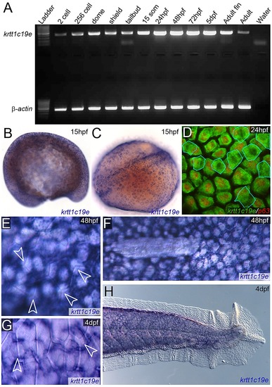
Timing of expression of krtt1c19e in the epidermis. A: RT-PCR of krtt1c19e (upper gel) at stages given and compared to β-actin positive control (lower gel) showing expression of krtt1c19e at all stages. Negative water control is given in far right lane. B–H: In situ hybridisations of krtt1c19e detected fluorescently (D) or by chromogenic precipitate (B–C, E–H) at 15 pf (B–C), 24 hpf (D), 48 hpf (E–F) and 4 dpf (G–H). Lateral (B) and dorsal (C) view of krtt1c19e in situ hybridisation at 15 hpf, when specific epidermal expression can be discerned. Counter-staining fluorescent in situ hybridisations with an antibody against ΔNp63 (red; D) demonstrates that in addition to the predominant basal keratinocyte expression, there is some low level expression of krtt1c19e in the EVL (outlined in blue) at 24 hpf. The predominant expression of krtt1c19e in basal layers at 48 hpf and 4 dpf is demonstrated through imaging the boundary of overlying EVL cells with Nomarski optics (arrowheads; E, G) and observing the lateral displacement of krtt1c19e expressing cells by the primordium at the end of its migration (F). The expression of krtt1c19e remains excluded from the epidermis of the medial fin at 4 dpf (H). |