
Fig. 1
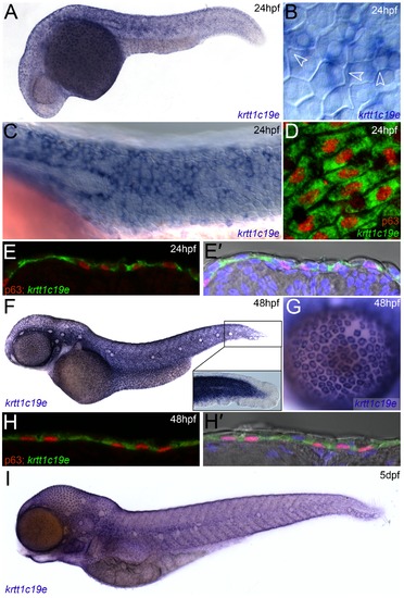
Expression of krtt1c19e in basal keratinocytes. In situ hybridisation of krtt1c19e at 24 hpf (A-E′), 48 hpf (F-H′) and 5 dpf (I), imaged laterally (A-D, F-G, I) or after cryosectioning (E-E′, H-H′). Overviews of embryos are shown at 24 hpf (A), 48 hpf (F) and 5 dpf (I), showing broad skin expression. Epidermal cells were visualised by counterstaining with DAPI (blue - E′, H′) or by Nomarski optics (B-C, F inset, E′, H′), and basal cell nuclei were immunolabelled using an antibody against ΔNp63 (red D-E′, H-H′). Strong krtt1c19e epidermal expression can be seen in the basal keratinocytes with the borders of overlying EVL cells intersecting basal cells (B - arrowheads). A gap in the krtt1c19e in situ signal is seen in the epidermis corresponding to the location of the migrating lateral line primordial (C). krtt1c19e expressing keratinocytes have ΔNp63 immunoreactive nuclei (D-E′, H-H′) and are seen below EVL cells in cryosections (E′, H′). A higher magnification of the tail region of the 48 hpf embryo is shown inset (F), with expression excluded from the fin epithelium, whilst a single layer of keratinocytes can be seen over the eye as part of the cornea (G). |
| Gene: | |
|---|---|
| Antibody: | |
| Fish: | |
| Anatomical Terms: | |
| Stage Range: | Prim-5 to Day 5 |