
Fig. 1
- ID
- ZDB-FIG-140319-29
- Publication
- Püschel et al., 1992 - Sequence and expression pattern of pax-6 are highly conserved between zebrafish and mice
- Other Figures
- All Figure Page
- Back to All Figure Page
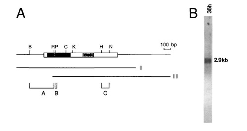
Structure and expression of pax-6. (A) Schematic representation of the pax-6 cDNAs. The box indicates the coding region, the darkly stippled regions the paired domain, the lightly stippled regions the homeodomain and the black region the 14 amino acid insert in the paired domain of zf pax-6b. Lines indicate the two classes of isolated cDNAs (I: n9, nl6, n27, nlO8, nl30; II: n8, nl5). The 5′ ends of class II clones were near position 500 of the sequence shown in Fig. 2. Clone nlO8 contains an additional sequence 5′ of the insert which most likely represents an intron (data not shown) which is not present in the other partially sequenced cDNA′s. Brackets indicate probes used for Northern blot hybridization and in situ hybridization. (A) a BamHl - EcoRI fragment, (B) a cloned oligonucleotide containing the 42 bp insert of zf pax-6b, (C) a Hindlll - Ndel fragment. (B) Northern blot of 36 h embryonic mRNA hybridized with a pax-6 probe. A Northern blot with 5 ng of poly(A)+ RNA was hybridized with probe A. A message of approximately 2.9 kb was detected. The same result was obtained when using probe C. (B) BamHI, (C) ClaI, (H) HindIII, (K) KpnI, (N) NdeI, (P) PstI, (R) EcoRI. |
| Gene: | |
|---|---|
| Fish: | |
| Anatomical Term: | |
| Stage: | Prim-25 |