
Fig. 4
- ID
- ZDB-FIG-140304-1
- Publication
- Decker et al., 2014 - Abnormal differentiation of dopaminergic neurons in zebrafish trpm7 mutant larvae impairs development of the motor pattern
- Other Figures
- All Figure Page
- Back to All Figure Page
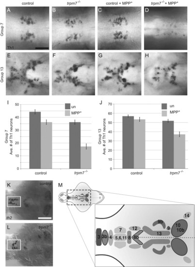
trpm7 mutants are sensitized to MPP+. (A–H) Dorsal views of 5-dpf larvae of the indicated genotype, treated with 500 μm MPP+ or vehicle control as indicated. Individual panels show distinct groups of Th1 IR cells in the following brain regions: (A–D) pretectal diencephalon (group 7); (E–H) periventricular hypothalamus (group 13); and I and J) bar chart showing average numbers of Th1 IR-positive cells in each group (n=11 animals scored). In the untreated samples, the number of Th1 IR cells was significantly lower in mutants than controls in group 7 (control avg. +/- SEM: 44 +/- 2, mutant: 36 +/- 2; Two way ANOVA with Bonferroni post-hoc test, p<0.001).. In group 13, (control: 56 +/- 1, mutant: 51 +/- 2) there was not a significant difference in the number of Th1 IR cells between untreated controls and untreated mutants. In both groups, MPP+ treatment effected a larger reduction in the number of Th1 IR cells in mutants than in controls (group 7, control+MPP+: 39 +/- 2, mutant+MPP+: 17 +/- 2; group 13, control+MPP+: 53 +/- 2, mutant+MPP+: 37 +/- 3). In group 7 the effect of MPP+ in mutants was almost three times larger than in controls, and in group 13 it was over four times larger; in both groups, mutants were significantly more sensitive to MPP+ treatment than controls (p<0.001). (K and L) Dorsal views of dissected brains from (K) control or (L) trpm7 mutant larvae, processed to reveal th2 mRNA. The pre-optic area (group 3b) is outlined in white. (th2-positive cells: control: 14 +/- 1, n=9; mutant: 7 +/- 5, n=12; p<0.001). Scale bar in A=50 μm, applies to A-H. Scale bar in K, =100 μm, applies to K,L. (M) Schematic illustrating the numbering scheme of Th1 and th2 positive (outlined in black) neuron clusters where darkness indicates more ventral groups (1 olfactory bulb; 2 subpallium; 3–4 preoptic area; 5,6,11 diencephalon; 7 pretectum; 8 anterior paraventricular organ; 9 interior paraventricular organ; 10 posterior paraventricular organ; 12 posterior tuberal nucleus; 13 hypothalamus; 14 locus coeruleus; 15-16 medulla oblongata; 17 area postrema; see Table S1 for additional details) |
| Genes: | |
|---|---|
| Fish: | |
| Condition: | |
| Anatomical Terms: | |
| Stage: | Day 5 |
Reprinted from Developmental Biology, 386(2), Decker, A.R., McNeill, M.S., Lambert, A.M., Overton, J.D., Chen, Y.C., Lorca, R.A., Johnson, N.A., Brockerhoff, S.E., Mohapatra, D.P., Macarthur, H., Panula, P., Masino, M.A., Runnels, L.W., and Cornell, R.A., Abnormal differentiation of dopaminergic neurons in zebrafish trpm7 mutant larvae impairs development of the motor pattern, 428-39, Copyright (2014) with permission from Elsevier. Full text @ Dev. Biol.