|
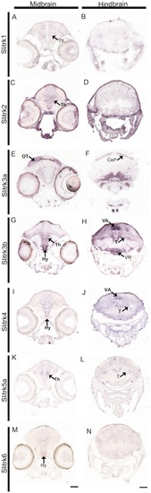
A-N Slitrk expression at 10 days post fertilization is observed in the developing midbrain and hindbrain. In situ hybridization of 10 dpf zebrafish sections demonstrates differential expression patterns of the seven expressed slitrks in the midbrain (A,C,E,G,I,K,M) and hindbrain (B,D,F,H,J,L,N). Multiple slitrks are expressed in the thalamus (A,C,G,I,K) and hypothalamus (G,I,M), and slitrk5b is detected in the optic tectum. Notably, slitrk3b is highly expressed in the cerebellar valvula and ventricular recess of the hypothalamus. CeP, cerebellar plate; Hy, hypothalamus; OT, optic tectum; T, tegmentum; Th, thalamus; VA, cerebellar valvula; VR, ventricular recess of hypothalamus. Scale bars, 50 μm.
|