
Fig. 2
- ID
- ZDB-FIG-131218-1
- Publication
- Phillips et al., 2013 - The cone-dominant retina and the inner ear of zebrafish express the ortholog of CLRN1, the causative gene of human Usher syndrome type 3A
- Other Figures
- All Figure Page
- Back to All Figure Page
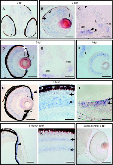
clrn1 is expressed in the retina, inner ear, and lateral line at embryonic, larval, and adult stages. A digoxigenin-labeled cRNA probe was used to detect clrn1 transcript on sectioned tissue from 2 dpf to 6 months of age. (A-C) Developing eyes and ears are enriched for clrn1 in the outer (closed arrow) and inner (open arrow) nuclear layers and ganglion cell layer (bracket) of the retina, (A and B), in mechanosensory hair cells (closed arrowhead in C) and supporting cells (open arrow in C) of the ear and in the neuromast cells (nm). (D-F) Retinal expression is retained through larval stages in all nuclear layers of the retina and in the proliferating cells of the ciliary marginal zone (open and closed arrows in D indicate inner nuclear layer and outer nuclear layer cells, respectively; bracket indicates the CMZ), all sensory patches of ear, and neuromasts (E). Additional expression is noted in brain (asterisk in F). 7 dpf larva in F was raised in PTU to suppress melanocyte formation. (G-I) 10 dpf zebrafish larvae express clrn1 in the anterior chamber of the retina (arrow in G) and continued expression is present in the photoreceptor layer (closed arrowhead in H) inner retina (open arrowhead in H) and CMZ (bracket in G). clrn1 expression is detected both in mechanosensory hair cells (closed arrowhead in I) and supporting cells (open arrow in I) of the sensory patches (anterior macula shown in I). (J and K) Continued expression of clrn1 transcript in the anterior chamber (open arrowhead in J), photoreceptors (closed arrow in K), and inner retinal cells (open arrow in K) of adult retinas. No signal is detected in tissues incubated with a sense probe (shown in L: 3 dpf larval retina treated with PTU). C,E,I: lateral views with anterior to the left; all others horizontal views, with anterior to the bottom. Abbreviations: am: anterior macula; lc: lateral crista; nm: neuromast. Scale bars: A-C, E, H, I and L: 20 µm. D, G, J and K: 50 µm. F: 100 µm. |
| Gene: | |
|---|---|
| Fish: | |
| Anatomical Terms: | |
| Stage Range: | Long-pec to Adult |
Reprinted from Gene expression patterns : GEP, 13(8), Phillips, J.B., Västinsalo, H., Wegner, J., Clément, A., Sankila, E.M., and Westerfield, M., The cone-dominant retina and the inner ear of zebrafish express the ortholog of CLRN1, the causative gene of human Usher syndrome type 3A, 473-81, Copyright (2013) with permission from Elsevier. Full text @ Gene Expr. Patterns