
Fig. S3
- ID
- ZDB-FIG-131029-54
- Publication
- Fang et al., 2013 - Translational profiling of cardiomyocytes identifies an early Jak1/Stat3 injury response required for zebrafish heart regeneration
- Other Figures
- All Figure Page
- Back to All Figure Page
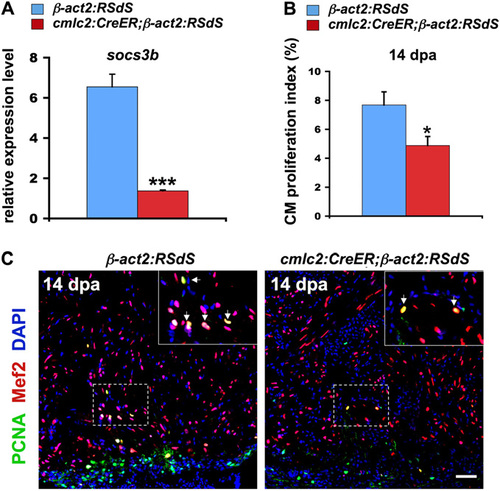
Inhibition of Stat3 signaling in transgenic fish. (A) qPCR indicating reduced expression of the Stat3 target gene socs3b in dnStat3-expressing (cmlc2: CreER; β-act2:RSdS) ventricles at 1 dpa, signifying that endogenous Stat3 activity was impaired. Data are mean ± SEM n = 3, ***P < 0.001, Student t test (unpaired, two-tailed). Expression levels were normalized to that of β-actin2, and further normalized to that of β-act2:RSdS. (B) cmlc2:CreER;β-act2:RSdS and control β-act2:RSdS fish were treated with 4-hydroxytamoxifen (4-HT) at 8 dpa to induce dnStat3 expression, and cardiomyocyte proliferation was analyzed in 14-dpa ventricles. There was an <36% reduction in cardiomyocyte proliferation indices after dnStat3-GFP expression. Data are mean ± SEM n = 5–7, *P < 0.05, Student t test (unpaired, two-tailed). (C) Confocal images indicating reduced cardiomyocyte proliferation in 14-dpa ventricles, after dnStat3-GFP induction at 8 dpa. (Insets) Higher-magnification images of the squares; arrows, proliferating cardiomyocytes. (Scale bar, 50 μm.) |
| Gene: | |
|---|---|
| Fish: | |
| Condition: | |
| Anatomical Term: | |
| Stage: | Adult |