
Fig. 3
- ID
- ZDB-FIG-130912-23
- Publication
- Tse et al., 2013 - Early embryogenesis in zebrafish is affected by bisphenol A exposure
- Other Figures
- All Figure Page
- Back to All Figure Page
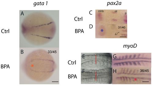
Bisphenol A alters somite formation at the 8–10 somite stage. Lateral expansion of the presumptive hematopoietic cell marker (gata 1) was indicated by an orange asterisk, which indicates the dorsalized phenotype in the bisphenol A (BPA)-exposed embryos (A,B). pax2a expression at the 8–10 somite stage, dorsal view (C,D). A blue asterisk indicates abnormal developmental pattern in the mid-hindbrain boundary (mhb) in BPA-exposed embryos. Additionally, an arrow marks the missing of the 2 otic vesicles in BPA-exposed embryos (D). Somite morphology of the control (E) and the BPA-exposed embryos (F) at the 8–10 somite stage. Lateral expansion of somite muscles was observed (red dotted lines). The somite marker, myoD, showed widened and diffused expression in the BPA-exposed embryos (red asterisk) as compared to the control (G,H). mhb, mid-hindbrain boundary; ot, otic vesicle; p, pronephric precursor expression domain. All were head to the left. Scale bars: 75μm (A,B); 200μm (C,D); 150μm (E–H). The number of embryos with the presented phenotype is shown in the top right corner of the panel. |