
Fig. 1
- ID
- ZDB-FIG-130808-10
- Publication
- Hayes et al., 2013 - Expression of glycosaminoglycan epitopes during zebrafish skeletogenesis
- Other Figures
- All Figure Page
- Back to All Figure Page
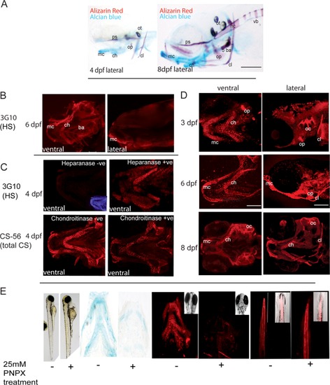
Chondroitin sulfate and Heparan sulfate are widely expressed in the developing zebrafish skeleton. A: Alcian blue- and alizarin red-stained skeletal preparations showing cartilage (blue) and mineralised tissue (red) at 4 and 8 dpf (lateral). B: Heparan sulfate labelling of the head with monoclonal antibody 3G10 at 6 dpf. C: Immunohistochemistry controls of 3G10 (labelling heparan sulphate) and CS56 (labelling native chondroitin sulphate), with and without chondroitinase ABC/heparitinase digestion as labelled at 4dpf. Heparitinase treatment is required to generate the epitope recognised by the 3G10 antibody, as such the heparitinase untreated fish show very limited immunoreactivity. CS56 antibody recognises a currently uncharacterised epitope present in native CS chains; treatment with chondroitinase ABC decreases immunoreactivity but doesn′t completely prevent antibody binding. Ventral views with anterior to top. Inset in left-most panel with low levels/no labelling of antibodies show DAPI stained or brightfield images for orientation. D: Chondroitin sulfate labelling of the head from 3–8 dpf with monoclonal antibody CS-56. E: Treatment of larvae with the GAG chain inhibitor PNPX leads to decreased GAG synthesis and decreased labelling with CS-56 in newly synthesised cartilage elements, demonstrating that CS-56 specifically labels CS chains. Images are all at 4dpf after treatment with PNPX (controls with DMSO) from 50 hpf. Left panels: Brightfield views of whole larvae to show that while morphology is relatively normal heart oedema is present. Second pair of panels: Flat-mounted cartilages of the ventral jaw stained with Alcian blue to reveal GAG content. Treatment with PNPX leads to significant reduction in cartilage GAG levels. Third pair of panels: Confocal stacks of the central jaw of zebrafish labelled with CS-56 antibody at 4dpf, treatment with PNPX leads to a significant reduction in cartilage labelling of CS-56 such that levels are comparable with the reduction in GAG synthesis observed by Alcian blue labelling. Right pair of panels: Tail of the zebrafish labelled with CS-56, comparable labelling of the notochord is seen following treatment with PNPX, likely because notochord synthesis of GAGs occurs between 24 and 48hpf prior to the onset of treatment with PNPX. Insets in images with low levels/no labelling of antibodies show DAPI stained or brightfield images for orientation. mc, Meckel′s cartilage; ch, ceratohyal; ba, branchial arches; op, operculum; ps, parasphenoid; oc, otic capsule; ot, otiliths; cl, cleithrum; 5ba, 5th branchial arch and teeth; nc, notochord; vb, developing vertebrae; ha, haemal arch; na, neural arch; sb, somite boundaries; +ve, positive; ve, negative. Anterior is to left in all images. Scale bars = 100 μm in all panels. |
| Antibodies: | |
|---|---|
| Fish: | |
| Condition: | |
| Anatomical Terms: | |
| Stage Range: | Protruding-mouth to Days 7-13 |