
Fig. 3
- ID
- ZDB-FIG-130719-4
- Publication
- Riera et al., 2013 - CERKL Knockdown Causes Retinal Degeneration in Zebrafish
- Other Figures
- All Figure Page
- Back to All Figure Page
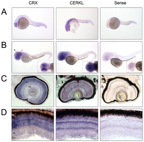
Cerkl in situ hybridization on embryo and adult zebrafish. (A and B) Whole-mount RNA in situ hybridization analysis showing cerkl expression in the retina and brain of embryos at 24 and 50 hpf. R, retina; L, lens. (C and D) In situ hybridization on zebrafish retina cryosections of 72 hpf embryos and adult tissue. Cerkl expression is detected in the three nuclear layers of the embryo retina, whereas adult expression appears in the inner segment of the photoreceptors and some cells located at the basal layer of the INL. Positive control (antisense CRX), strongly labels the inner photoreceptor segment and inner nuclear layer. The negative control was performed with sense cerkl. RPE, retinal pigment epithelium; PR, photoreceptor; ONL, outer nuclear layer; OPL, outer plexiform layer; INL, inner nuclear layer; IPL, inner plexiform layer; GCL, ganglion cell layer. |
| Gene: | |
|---|---|
| Fish: | |
| Anatomical Terms: | |
| Stage Range: | Prim-5 to Adult |