
Fig. 6
- ID
- ZDB-FIG-130719-6
- Publication
- Riera et al., 2013 - CERKL Knockdown Causes Retinal Degeneration in Zebrafish
- Other Figures
- All Figure Page
- Back to All Figure Page
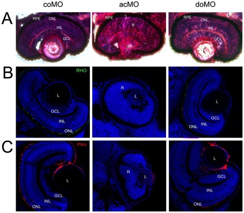
Eye histology in control and ZFcerkl morpholino-injected embryos at 72 hpf. (A) Zebrafish eye sections were stained with H&E. Control morphants (coMO) showed normal retinal lamination with three cell layers (GCL, INL, and ONL). In “mild” acMO-injected embryos, lamination did not occur and the three layers were not visible. The retinal pigment epithelium (RPE) developed normally in control and ZFcerkl morphants. (B) Immunostaining with anti-rhodopsin and (C) anti-PNA identified rod and cone outer segments, respectively, in control and doMO morphants. Outer segments were absent in acMO morphants. Nuclei were stained with DAPI. Some nonspecific staining was seen in the lens when stained with PNA. Photographs were at×40 magnification. |
| Fish: | |
|---|---|
| Knockdown Reagent: | |
| Observed In: | |
| Stage: | Protruding-mouth |