
Fig. S3
- ID
- ZDB-FIG-130625-37
- Publication
- Schmid et al., 2013 - Loss of ALS-associated TDP-43 in zebrafish causes muscle degeneration, vascular dysfunction, and reduced motor neuron axon outgrowth
- Other Figures
- All Figure Page
- Back to All Figure Page
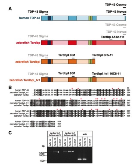
Schematic representation of antibody binding sites, alignment of C termini of human TDP-43, Tarbpl_tv1, and Tardbp, and up-regulation of tardbpl_tv1 mRNA upon loss of Tardbp. (A) Epitopes of respective antibodies (written in black) are indicated as a black line above the schematically represented proteins (protein domains as in Fig. 1). Lines and antibody names in gray indicate cross-reactivity of antibodies raised against human TDP-43 protein with the corresponding epitopes of zebrafish homologs. (B) Amino acid alignment of exon 5 of Tardbpl_tv1 with the corresponding sequence of zebrafish Tardbp and human TDP-43 (identical amino acids are not highlighted, identical amino acids in two of the three amino acids are highlighted in light gray and nonidentical amino acids are highlighted in dark gray). Amino acids mutated in ALS/FTLD patients are marked with a red asterisk. (C) Semiquantitative RT-PCR with primers specific for tardbpl_tv1 (tardbpl_tv1 primer pair 1 and 2) and actin as a loading control. Each primer pair is used on cDNA generated from buffer injected control embryos, Tardbp knockdown embryos, or a negative control with no cDNA added, respectively. Arrowheads indicate the up-regulation of tardbpl_tv1 transcript upon Tardbp KD compared with buffer injected control embryos. |