|
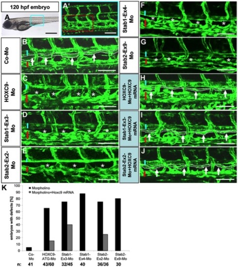
Silencing of HOXC9, Stab1 and Stab2 expression in zebrafish inhibits formation of the thoracic duct (TD).
(A) 120 hpf tg(fli1:EGFP) zebrafish embryo. Blue box marks the region magnified in (A2). (A2) Trunk vasculature of the embryo shown in (A). Red box marks the region magnified in (B–J). (B) Normal formation of the TD (arrows and dotted line) in 120 hpf tg(fli1:EGFP) zebrafish embryos after injection of control morpholino. Blue bar marks dorsal aorta and red bar marks cardinal vein. (C–G) Silencing of HOXC9, Stab1 and Stab2 expression using the indicated morpholinos disrupted the formation of the TD (asterisks) in 120 hpf tg(fli1:EGFP) zebrafish embryos. (H–J) Co-injection of 50 pg HOXC9 mRNA rescued the defects in TD formation caused by silencing of HOXC9, Stab1 and Stab2 using the indicated morpholinos. (K) Quantification of defects in TD formation of embryos shown in (A–J) including rescue experiments with 50 pg HOXC9-mRNA. Black scale bar: 500 μm. White scale bar: 50 μm.
|