
Fig. 1
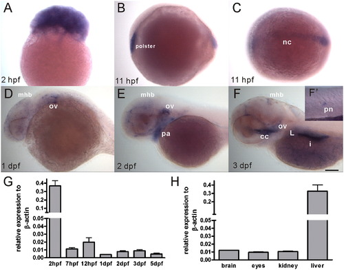
Spatiotemporal expression of the manf transcript during zebrafish embryogenesis and in adult organs. (A–F) manf expression pattern was revealed by whole-mount in situ hybridization, anterior to the left. (A) lateral view of a 2 hpf embryo, (B) lateral view of a 11 hpf embryo, (C) dorsal view of a 11 hpf embryo, (D–F) lateral view of a one dpf, two dpf and three dpf embryo, (F′) high magnification view of a posterior neuromast. (G) manf mRNA levels were analyzed by quantitative PCR. Thirty embryos/group were collected at defined stages. Each experiment was repeated three times using independent RNA isolations. (H) Adult tissues were collected from five individual one-year old male fish and analyzed by qPCR. cc, cranial cavity; i, intestine; L, liver; mhb, midbrain–hindbrain boundary; nc, notochord; ov, otic vesicles; pa, pharyngeal arches; pn, posterior neuromast. Scale bar=100 μm. |
| Gene: | |
|---|---|
| Fish: | |
| Anatomical Terms: | |
| Stage Range: | 64-cell to Adult |
Reprinted from Developmental Biology, 370(2), Chen, Y.C., Sundvik, M., Rozov, S., Priyadarshini, M., and Panula, P., MANF regulates dopaminergic neuron development in larval zebrafish, 237-249, Copyright (2012) with permission from Elsevier. Full text @ Dev. Biol.