
Fig. 4
- ID
- ZDB-FIG-120725-43
- Publication
- Vasilyev et al., 2012 - Mechanical Stretch and PI3K Signaling Link Cell Migration and Proliferation to Coordinate Epithelial Tubule Morphogenesis in the Zebrafish Pronephros
- Other Figures
- All Figure Page
- Back to All Figure Page
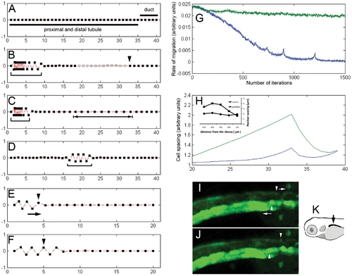
Modeling pronephric migration and proliferation. (A) Initial arrangement of cells in the simulation. Squares: cell positions. All cells in the chain are presumed to be responsive to fluid flow. The difference between the tubule segment (long bar) and the duct segment (short bar) is that cells in the tubule segment migrate faster than cells in the duct segment. (B) Collective migration results in piling up of epithelial cells in the proximal nephron (bracket) and an increase in total number of epithelial cells (gray squares indicate newly formed cells) due to stretch-dependent distal tubule proliferation. The arrowhead points to the zone of proliferation. The number of iterations = 700. (C) When the threshold for stretch-induced cell proliferation was increased, migration came to a premature halt and, as a result, proximal convolution was significantly reduced (bracket) and distal tubule became overstretched (bracketed arrow). The number of iterations = 700. (D) When only the distal half of the kidney is subjected to the directional migration bias, the ectopic convolution develops (bracket). The number of iterations = 100.The starting number of cells in simulations A–D was 40. (E,F) The model predicted that if at any time during active migration (such as in E (100 iterations)) the cue for directional migration (fluid flow) was eliminated, the direction of migration would temporarily reverse (F, arrow in E). (E, F) Arrowheads point to the same cell. The additional number of iterations in F = 100. The starting number of cells in simulations E–F was 20. (G) Simulations predicted that inhibiting distal proliferation should result in premature arrest of migration (lower trace) while in control condition the migration rate remained relatively constant (upper trace). The migration rate was measured over the five cells (20th–24th cells counting from the back end of the chain). (H) Inhibiting distal proliferation in live embryos using LY294002 resulted in linear stretch of the distal kidney epithelial cells as evidenced by the increase in the internuclear distance (inset). The same effect is predicted in our simulations (upper trace – no proliferation condition vs. lower trace – control condition). The total number of cells in this simulation = 40. The distal half is displayed. (I,J) Reversal of the direction of migration can be observed during stochastic transient tubule obstruction. (I) Transiently obstructed tubule (above) and unobstructed tubule (below) in the ET33d10 transgenic fish at time 0. (J) the same two tubules 1 hour later. Arrowheads point to the individual traced cell in each tubule. Arrows indicate the direction of migration. See also Movie S10. (K) Diagram showing the portion of the zebrafish kidney that was imaged in I and J. |