
Fig. S2
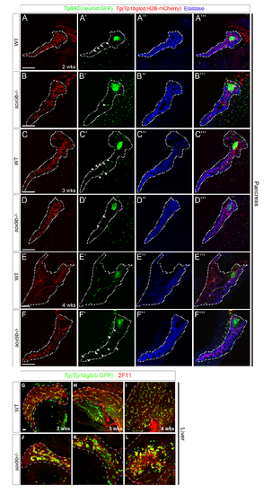
Time course analyses of the pancreas (A–F) and liver (G–L) in wild-type and sox9b mutant larvae at 2, 3, and 4 weeks. (A–F) Tg(Tp1bglob:H2B-mCherry);TgBAC(neurod:GF P) double transgenic fish from sox9b heterozygote incrosses were raised together and 40 of them were fixed and genotyped at each time point. The pancreas, along with the gut, of 6 to 11 wild-type or sox9b homozygous mutant fish were dissected, stained with anti-dsRed (red), anti-GFP (green) and anti-Elastase (blue) antibodies and mounted for confocal imaging. Whereas wild-type pancreata showed complex intrapancreatic duct networks at 2 and 3 weeks of age with several main pancreatic ducts (A, C), sox9b mutant pancreata failed to expand and ductal cells stayed in clusters (B, D). At 4 weeks of age, the morphological differences between wild-type and sox9b mutant pancreata were even more obvious: wild-type pancreata spread over the gut starting to form lobes whereas sox9b mutant pancreata were still primitive in appearance (E, F). The defect in pancreatic growth in sox9b mutant fish is associated with a global growth retardation of the fish as indicated by the smaller size of the fish and the frequent occurrence of an unlooped gut at 4 weeks (data not shown). In addition to pancreatic duct morphological defects, sox9b mutant fish showed a deficiency in secondary islet formation as assessed by TgBAC(neurod:GFP) expression (arrowheads in B2, D2, F2). However, at the equivalent stages, wild-type fish exhibited multiple clusters of TgBAC(neurod:GFP)-positive cells along the intrapancreatic ducts (arrowheads in A2, C2). All images are projections of confocal z-stacks. Ventral views, anterior to the top right. Dashed lines delineate the pancreas. Scale bars, 100 μm. (G–L) Wild-type and mutant larvae were sorted at 5 dpf based on their intrahepatic ductal system phenotypes (as assessed by the pattern of Tg(Tp1bglob:GFP) expression) and raised separately. At 2 (G, J), 3 (H, K), and 4 weeks (I, L) of age, 2 wild-type and 2 mutant fish were fixed for immunostaining. Cryosections were stained with 2F11 (red) and anti-GFP (green) antibodies. In the liver, whereas the intrahepatic biliary network continued to expand between 2 and 4 weeks in wild-types (G–I), the biliary cells in sox9b mutants remained clustered and never assumed normal morphogenesis (J–L). Moreover, the expression of Tg(Tp1bglob:GFP) largely overlapped with 2F11 labeling in wild-type livers (G–I). In contrast, in sox9b mutant livers, we observed an increasing number of cells that were labeled by 2F11, but did not express Tg(Tp1bglob:GFP) (J–L). These cells formed large clusters by 4 weeks of age (L). All images are projections of confocal z-stacks. Sagittal sections, anterior to the top. Dashed lines in (G–L) delineate the liver. Li, liver; Pa, pancreas. Scale bar, 20 μm. |