
Fig. 2
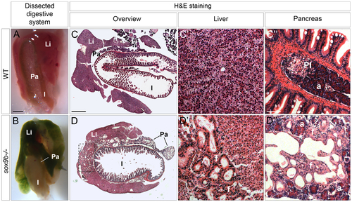
Adult sox9b mutants develop cholestasis associated with fibrosis, duct proliferation, and dilation in both the liver and pancreas. (A–B) Dissection of the digestive system of 5 month-old wild-type (A) and sox9b homozygous mutant (B) fish reveals a green mutant liver and pancreas reflecting the accumulation of bile in both organs. Anterior to the top. (C–D) Hematoxylin-and-eosin (H&E) staining of histological cross-sections of wild-type (C) and sox9b mutant (D) digestive tracts shows abnormal duct morphology in the mutant liver and pancreas. These duct malformations are focalized around the connection with the extrahepatic duct (dashed rectangle in D) in the liver, whereas they are spread over the entire pancreas. Higher magnifications of liver (C′–D′) and pancreas (C′′–D′′) reveal dilated ducts surrounded by fibrotic tissue (pink staining in D3 labeled as “f”) in both organs in sox9b mutants. Li, liver; Pa, pancreas; I, intestine; PI, primary islet; a, acinar compartment. Scale bars, 1 mm in A–B; 500 μm in C–D; 50 μm in C′–D′′. |
| Fish: | |
|---|---|
| Observed In: | |
| Stage: | Adult |