|
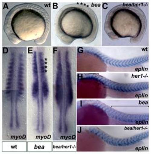
Analysis of the her1hu2124/bea tm98 double mutant phenotype. Brightfield images of wild type, beatm98 and her1hu2124/beatm98 mutant embryos at the 10–12 somite stage, lateral views, anterior to left. Compared to the wild type embryo (A), the somite borders posterior of the 4th somite are disrupted in the beatm98 mutant (B, asterisks indicate correctly formed somites). All somitic borders are disrupted in the her1hu2124/beatm98 double mutant (C). In situ hybridisation analysis of myoD expression at 10–12 somites (D–F), dorsal views, anterior to top. In line with the morphological phenotypes, half segmental myoD expression is disrupted posterior to the 4th somite in beatm98 (E, asterisks mark residual expression in somites 1–4) and along the entire body axis in her1hu2124/beatm98 double mutants (F) compared to wild type (D). In situ analysis of eplin expression at prim 6 stage (G–J) lateral views, anterior to left. eplin is expressed in v-shape at the somite borders in the wild-type (G). Disturbed eplin expression is observed in the first somites of the her1hu2124 mutant (H, bracket), posterior to the somite 4 in the beatm98 mutant (I, bracket) and in all somites in the double mutant situation (J). (A–F) 10–12 somite stage, (G–J) prim 6 stage.
|