
FIGURE
Fig. 6
- ID
- ZDB-FIG-120525-7
- Publication
- Mattes et al., 2012 - Wnt3 and Wnt3a are required for induction of the mid-diencephalic organizer in the caudal forebrain
- Other Figures
- All Figure Page
- Back to All Figure Page
Fig. 6
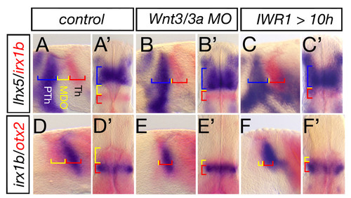
Blockage of Wnt signaling lead to lack of MDO tissue. Lateral views and dorsal views (marked by ′) of embryonic heads at 28 hpf. Wnt3/wnt3a morphant embryos lack the organizer tissue and the lhx5 positive prethalamus (PTh, blue) abuts the irx1b thalamus (Th, red) (A-B′, n = 54/78). A similar phenotype is observed in embryos treated with 30 μM IWR1 from 10 to 28 h to inhibit canonical Wnt signaling (n = 15/17). Hence, the otx2 positive MDO (yellow) is lacking if Wnt3/Wnt3a function is knocked-down (n = 44/80, D-E′), likewise after inhibition of canonical Wnt signaling (F, F′, n = 14/24). |
Expression Data
| Genes: | |
|---|---|
| Fish: | |
| Condition: | |
| Knockdown Reagents: | |
| Anatomical Terms: | |
| Stage: | Prim-5 |
Expression Detail
Antibody Labeling
Phenotype Data
| Fish: | |
|---|---|
| Condition: | |
| Knockdown Reagents: | |
| Observed In: | |
| Stage: | Prim-5 |
Phenotype Detail
Acknowledgments
This image is the copyrighted work of the attributed author or publisher, and
ZFIN has permission only to display this image to its users.
Additional permissions should be obtained from the applicable author or publisher of the image.
Full text @ Neural Dev.