
Fig. 3
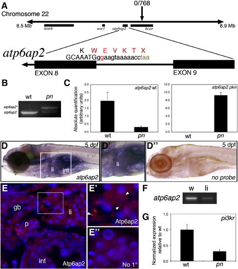
Mapping and genetic characterization of pn. (A) Schematic of mapping around the pn locus. A region of interest on chromosome 22 was identified as depicted, with a zero recombinant marker in the bcor gene. A splice donor site mutation following exon 8 of atp6ap2 was detected in pn mutants, which would result in the depicted sequence. (B) PCR of the wild-type (wt) and pn splice products, demonstrating the presence of the higher band (atp6ap2*), corresponding to the read-through product. (C) Quantitative PCR of the wt and pn atp6ap2 splice products, demonstrating a 10 × reduction in the wt product in pn, and a very large increase in the amount of the mutant transcript in pn. (D) In situ hybridization of atp6ap2 at 5 dpf demonstrates liver (“li”) and intestinal (“int”) expression of atp6ap2, shown in higher magnification in D2. Compare to (D3), which is a similarly processed 5 dpf larva without atp6ap2 riboprobe, with no specific staining. (E) Immunostaining of Atp6ap2 in 5 dpf cross-sections demonstrates staining in liver (“li”), intestine (“int”), pancreas (“p”), and gallbladder (“gb”). The area within the white rectangle is shown in E2, demonstrating punctate staining (white arrowheads), consistent with a vesicular localization, within the liver. (E3) Liver section from a 5 dpf larva processed similarly but without primary antibody, at the same magnification as (E2), showing no punctate staining. (E), (E2) and (E3) are counterstained with DAPI to show nuclei. (F) PCR of atp6ap2 from 5 dpf whole larvae (w) and from livers (li) isolated from 5 dpf larvae, showing expression of atp6ap2. (G) Quantitative PCR demonstrating a 5 × decrease in pi3kr1 expression, a gene regulated by Atp6ap2 via PLZF. |
| Genes: | |
|---|---|
| Antibody: | |
| Fish: | |
| Anatomical Terms: | |
| Stage: | Day 5 |
Reprinted from Developmental Biology, 365(2), Eauclaire, S.F., Cui, S., Ma, L., Matous, J., Marlow, F.L., Gupta, T., Burgess, H.A., Abrams, E.W., Kapp, L.D., Granato, M., Mullins, M.C., and Matthews, R.P., Mutations in vacuolar H(+)-ATPase subunits lead to biliary developmental defects in zebrafish, 434-444, Copyright (2012) with permission from Elsevier. Full text @ Dev. Biol.