
FIGURE
Fig. S2
Fig. S2
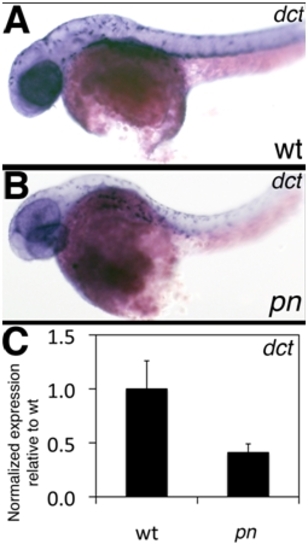
Expression of dct in pn. (A–B) Left lateral views of in situ hybridizations of 2 dpf wt (A) and pn (B) larvae for dct, a marker of melanocytes. Note that the wt and pn expression patterns are similar. (C) QPCR of dct expression at 5 dpf, showing a decrease in dct expression in pn. |
Expression Data
Expression Detail
Antibody Labeling
Phenotype Data
Phenotype Detail
Acknowledgments
This image is the copyrighted work of the attributed author or publisher, and
ZFIN has permission only to display this image to its users.
Additional permissions should be obtained from the applicable author or publisher of the image.
Reprinted from Developmental Biology, 365(2), Eauclaire, S.F., Cui, S., Ma, L., Matous, J., Marlow, F.L., Gupta, T., Burgess, H.A., Abrams, E.W., Kapp, L.D., Granato, M., Mullins, M.C., and Matthews, R.P., Mutations in vacuolar H(+)-ATPase subunits lead to biliary developmental defects in zebrafish, 434-444, Copyright (2012) with permission from Elsevier. Full text @ Dev. Biol.