|
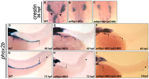
Enteric nervous system of arl6ip1 morphants is degenerated. Wild-type (WT) embryos (A, D, G), arl6ip1-MO1-injected embryos (B, E, H), and arl6ip1-MO1/p53-MO-injected embryos (C, F, I) were observed under dorsal view at 36-hpf (A-C) and under lateral view at 48 hpf (D, E, F) and 72 hpf (G, H, I). Neural crest cells (NCCs), which were labeled by crestin probe (arrowheads in A), of WT embryos exited the vagal region and migrated to the enteric region. However, the crestin-labeled NCCs of arl6ip1-MO1-injected and arl6ip1-MO1/p53-MO-injected embryos remained in the vagal region (B, C). Compared to WT embryos, either the arl6ip1-MO1-injected embryos or arl6ip1-MO1/p53-MO-injected embryos exhibited shortened enteric neurons and delayed migration (D vs. E; D vs. F; distance between two arrows). Additionally, the phox2b-positive enteric neurons were distributed throughout the entire gut to the anus (G). Arrows indicate the most posterior region of migration, and the asterisks represent the anus. The enteric neurons of arl6ip1-MO1-injected embryos and arl6ip1-MO1/p53-MO-injected embryos all failed to reach as far as the anus (H, I).
|