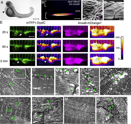
Anxa6/Dysf Double Morphant, Anxa6/Dysf Coexpression, and Ultrastructural Characterization of Muscle Damage
(A) Anxa6 and dysf (dysf i36e37) double morphants have curved body and severe cardiac edema (arrowhead) at 72 hpf.
(B) Muscle birefringence is almost completely absent.
(C) Misalignment (asterisks) and gaps between myofibers are visible by actin staining in the double morphants.
(D) Actin staining demonstrates normal myofiber alignment in uninjected control larvae.
(E) The accumulation of mTFP1-Dysf-coated membrane at the lesion (arrow) is rapid and coincides with Anxa6-mOrange1 appearance at the damaged sarcolemma. Increased fluorescence is depicted by lighter colors and higher surface plot profile. Note that the initial accumulation appears at the edges of the sarcolemma, from where it spreads over the damaged area, which is eventually sealed (arrowhead).
(F–N) Electron microscopy of 72 hpf myofibers. In (F), veratridine treatment leads to vesicle (arrowhead) accumulations under the sarcolemma in WT myofiber. In (G), high-magnification is shown of the boxed area in (F). In (H), cytoplasmic vesicles (arrowhead) often arise close to T-tubules (arrow). In (I), membrane blebbing and vesicles (arrowheads) could be observed at sites of fiber rupture. In (J), veratridine treatment leads to extensive muscle damage in anxa6 morphants. Large vacuolar structures (arrowheads) can be seen around ruptured myofibers. In (K), veratridine treatment enhances muscle damage in dysf i36e37 morphants. In some areas, correct sarcomeric filament alignment was evident (C2), whereas in other areas, it was significantly impaired (C1) or completely absent (C3).
(L) High-magnification image of veratridine-treated dysf i36e37 morphant muscle demonstrates misaligned sarcomeres, accumulation of vesicles (arrowhead) in the cytosol and T-tubule damage (arrows). The insert shows a magnified area (1 × 1 μm).
(M and N) Anxa6 and dysf double morphants have severe muscle damage in untreated (M) and veratridine-treated (N) conditions. Membrane blebbing and vesicles (arrowheads) could be detected at sites of cell rupture after veratridine treatment. Orientation of embryos in (A–D): anterior left, dorsal up.
Scale bars represent 500 μm (A and B); 20 μm (C and D); 1.6 μm (E); 2 μm (F, G, and J–M); 1 μm (H, I, and N).
|
